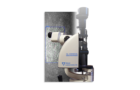
The HAI CL-1000eva Endothelium Viewing Attachment converts any compatible slit lamp into a specular microscope, providing a high-magnification view of endothelial cells with software that performs automatic image capture, frame selection, enhancement and analysis.
Features
- Live image of endothelium
- Optical pachymeter
- Auto selection of good endothelial images
- Analysis includes up to 200 cells
- Cell density, cell area and morphology
Description
Compact design
EVA’s small footprint saves valuable exam lane space while adding important clinical insight to your practice.
Fast and automatic cell analysis
EVA captures the best images so you can review and select a region of interest for analysis. The fully automated software calculates cell density, polymegathism (CV), pleomorphism (hex %) and corneal thickness within seconds.
Master-level control at your fingertips
EVA gives you total freedom to scan the patient’s cornea in every direction while seeing magnified endothelial cells in real-time on your monitor.
See more conditions
The powerful combination of live, manual tracking and auto camera trigger allows you to see even the more difficult clinical cases, like Fuch’s dystrophy, keratoconus and post-DSAEK surgery.
Additional Information
Type
Non-contact objective lens
Field Of View
250µm x 400µm
Camera
Monochrome CCD
Illumination
From slit lamp
Data Transfer
IEEE 1394 (FireWire) to PC
Power Input
110/220v – 60/50Hz
Weight
2.2lb (1.0kg)


